
Peering into the Nanoscale: AFM Images Bacteria, Nanoprisms, and More
TL;DR
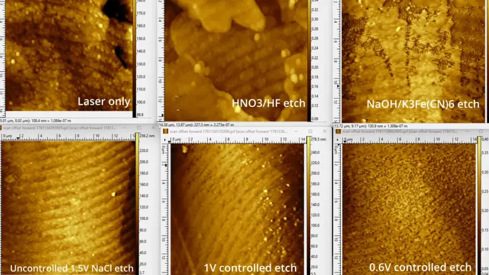
An atomic force microscope images surfaces by a tiny probe rather than light, achieving sub-nanometer detail; Ben Krasnow’s video shows the probe in action, then uses AFM to image nattō bacteria on a gelatin-coated silicon substrate to immobilize and orient them, and also captures silver nanoprisms, track-etched membranes, and laser-etched diffraction gratings, while noting ongoing work on etching methods and the existence of DIY AFMs.
Topics:sciencetechnology#afm#atomic-force-microscope#bacteria#diffraction-gratings#silver-nanoprisms#technology
Want the full story? Read the original reporting
Read on Hackaday