
New Ultrasound Method Tracks Glioblastoma’s Structural Manipulation of Brain Tissue in Awake Mice
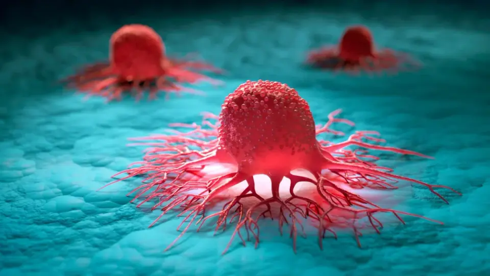
Researchers at Caltech have developed a trimodal ultrasound technique to observe glioblastoma growth in real-time within the brains of awake mice. By engineering tumor cells to produce gas vesicles, the team could track how the cancer displaces healthy tissue and remodels blood vessels over an 11-day period. This non-invasive method offers a new way to study the physical interactions between tumors and the brain, potentially aiding future treatment strategies.
Key points
- A team led by chemical engineer Mikhail Shapiro at Caltech published a paper in iScience detailing a new ultrasound imaging platform for studying neuro-oncological processes.
- The technique combines three modes: gas vesicle imaging to visualize the tumor, blood volume tracking for brain activity, and microbubble injection for detailed vascular mapping.
- The study involved implanting engineered human glioblastoma cells into the left thalamus of three mice and monitoring them over 11 days while the animals remained awake.
- Observations showed the tumor physically pushing the lateral geniculate nucleus out of position and altering local blood flow, with some vessels narrowing or becoming obstructed.
- The tumor exhibited its own rhythmic blood flow patterns, distinct from the coordinated patterns of the surrounding healthy brain tissue.
- The researchers emphasize this is a proof-of-concept study with a small sample size, and the glioblastoma model does not fully replicate the complexity of human cancer.
Background
This research builds on recent efforts to understand brain cancer mechanisms, such as the discovery of skull immune hubs that may improve anti-tumor responses in glioma models. It also follows broader trends in cancer research, including studies on alcohol-linked cancer risks and the protective effects of HPV vaccines against head and neck cancers. Additionally, it highlights the ongoing challenges in accessing treatments for rare brain cancers, as seen in recent cases involving denied insurance coverage for experimental therapies.
Why it matters
Glioblastoma has a 5-year survival rate of only 5 to 7 percent, largely because it intertwines with healthy brain tissue and blood vessels, making treatment difficult. Current imaging methods like MRI or optical imaging have limitations in capturing the full scope of tumor progression in awake animals. This new ultrasound technique provides a non-invasive way to observe how tumors manipulate their environment in real-time, which could lead to better understanding of tumor behavior and the development of more effective treatments that target the tumor's structural interactions with the brain.
What to watch
The next steps involve testing the technique on larger numbers of animals and using more realistic brain cancer models to verify if the observed patterns hold up. Researchers also plan to use the method to monitor how the brain and blood vessels respond to treatments, not just whether the tumor shrinks. Ultimately, the goal is to use this detailed observation to develop strategies to dismantle the tumor's 'nest' within the brain.
Want the full story? Read the original reporting
Read on ScienceAlert