
Histology-based tissue clocks reveal organ-specific aging patterns and disease signals
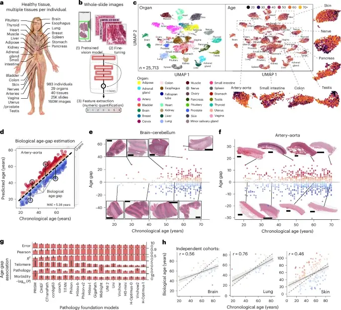
A large-scale study analyzes 25,713 whole-slide pathology images from 983 GTEx donors across 40 tissues to build tissue-specific “tissue clocks” that predict biological age from tissue morphology. The clocks achieve a mean absolute error of about 4.9 years and an r2 of ~0.69, with age gaps correlating to telomere shortening and subclinical pathologies. Higher age gaps accompany tissue-specific disease features and comorbidities, and tissue clocks generalize to independent brain, lung and skin cohorts. Comparisons with DNA methylation clocks show modality-specific strengths, while integrating RNA-seq data reveals tissue-specific gene expression changes linked to aging. Vision-language models help interpret aging-related histological features, and the authors demonstrate potential to predict tissue age gaps from blood expression, enabling non-invasive aging assessment and disease risk signals for conditions such as Alzheimer’s disease, stroke and Crohn’s disease.
- Histological aging signatures for monitoring tissue-specific aging and disease Nature
- Organs age at different speeds: A blood test might soon tell which ones medicalxpress.com
- AI ‘Tissue Clocks’ Reveal the Age of Individual Organs Inside Precision Medicine
- Blood test could soon reveal which organs are aging fastest Bioengineer.org
- #78: Medicine is treating your age, not your body – How organ-aging clocks could predict trouble years befor The Times of India
Want the full story? Read the original reporting
Read on Nature