
New AI tool aligns 3D scans with X-rays in seconds for surgery
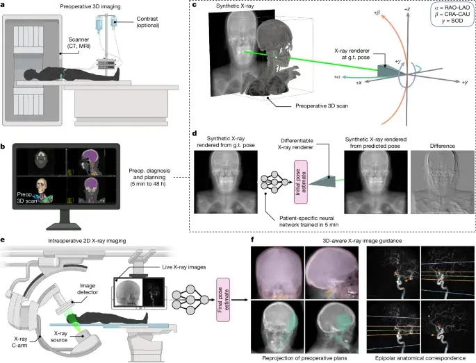
Researchers have developed a new open-source software called xvr that uses patient-specific neural networks to align 3D preoperative scans with 2D intraoperative X-rays in seconds. This method improves accuracy by an order of magnitude compared to existing tools and requires only five minutes of fine-tuning per patient.
Key points
- xvr is a self-supervised framework that combines patient-specific neural networks with gradient-based optimization for 2D/3D image registration.
- The system uses physics-based simulation to generate training data from a patient's own preoperative scan, eliminating the need for manual annotation.
- A foundation model was pretrained on thousands of whole-body scans, allowing for rapid patient-specific adaptation to any anatomical region.
- The tool achieves high accuracy in seconds across diverse anatomical structures and imaging modalities, outperforming existing methods by a factor of ten.
- The software is available open-source to make pan-anatomical registration accessible to broad clinical and research communities.
Background
This development builds on previous advancements in medical AI, such as the ARPA-H funding for patient-specific RNA therapies and breakthroughs in brain-computer interfaces. It addresses longstanding challenges in image-guided interventions where existing methods often require extensive manual labeling or fail to generalize across different anatomies.
Why it matters
Accurate alignment of 3D preoperative volumes with 2D intraoperative images is critical for the precision of surgical robotics and image-guided interventions. The new tool significantly reduces the time and complexity required for this process, potentially improving patient outcomes and expanding the accessibility of advanced imaging techniques in clinical settings.
What to watch
The open-source release of xvr is expected to facilitate broader adoption in clinical and research settings. Future developments may focus on integrating the tool into existing surgical workflows and expanding its application to other imaging modalities and anatomical regions.
Want the full story? Read the original reporting
Read on Nature