
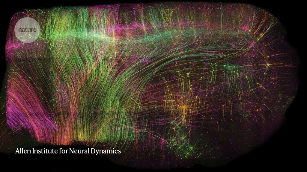
ExA-SPIM microscope revolutionizes brain imaging with high resolution and large samples
Scientists have developed a powerful microscope called ExA-SPIM (expansion assisted selective plane illumination microscopy) that allows for high-resolution imaging of large brain tissue samples without the need for slicing and reconstruction. By chemically removing lipids and expanding the tissue, researchers were able to image whole mouse brains at a resolution comparable to confocal microscopy. The approach, which can image an entire mouse brain in under a day, offers a faster and more accurate alternative to other whole-brain imaging techniques. The microscope design is open-source, but further development and documentation are needed to make it more accessible to other researchers.