A photographer’s ongoing project magnifies individual sand grains from beaches worldwide, revealing a surprising range of shapes and colors—fragments of volcanic rock, coral, shells, and microfossils—turning beach debris into a tiny, global museum of Earth’s geology and marine life.
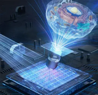
Researchers from the Smart Computational Imaging Laboratory (SCILab) at Nanjing University of Science and Technology unveil PCA-iSIM, a compact DMD-based incoherent structured-illumination microscopy framework that couples high-modulation coefficient mapping with principal component analysis to recover high-frequency details from low-contrast patterns, enabling real-time imaging at up to 30 frames per second with about 100 nm resolution and a ~70% simpler optical design than laser-based SIM; demonstrated in living cells imaging mitochondrial activity under challenging noise and setting changes.
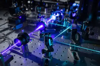
Researchers at the University of Tokyo developed pump-field-probe fluorescence microscopy that combines synchronized pulsed light with nanosecond magnetic pulses to detect spin-dependent, non-emissive intermediates invisible to standard fluorescence. Validated on flavin-based model systems, the method measures lifetimes and magnetic responses at biologically relevant, low concentrations and can operate with minimal sample damage, enabling time-resolved studies of short-lived dark states and offering potential for live-cell applications. This approach bridges fluorescence microscopy with spin chemistry, contributing to quantum biology and noninvasive diagnostics, with future plans to tackle more complex biological environments and separate overlapping reaction pathways.
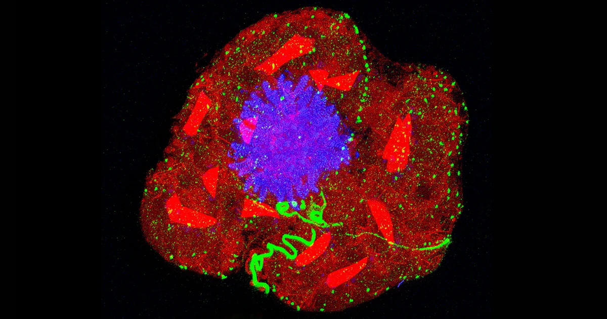
Expansion microscopy uses a diaper-inspired hydrogel to physically swell biological samples, enabling higher-resolution visualization of tiny cellular structures with standard microscopes. By improving dye penetration and preserving overall architecture, it democratizes microscopy and reveals detailed cytoskeletal diversity across species.
MIT researchers developed a terahertz microscope that uses spintronic emitters and a Bragg mirror to compress terahertz light to micron-scale spots, enabling imaging of the collective terahertz oscillations of superconducting electrons in BSCCO at cryogenic temperatures and overcoming the diffraction limit—a breakthrough for studying quantum modes in materials with potential implications for room-temperature superconductivity and terahertz devices.
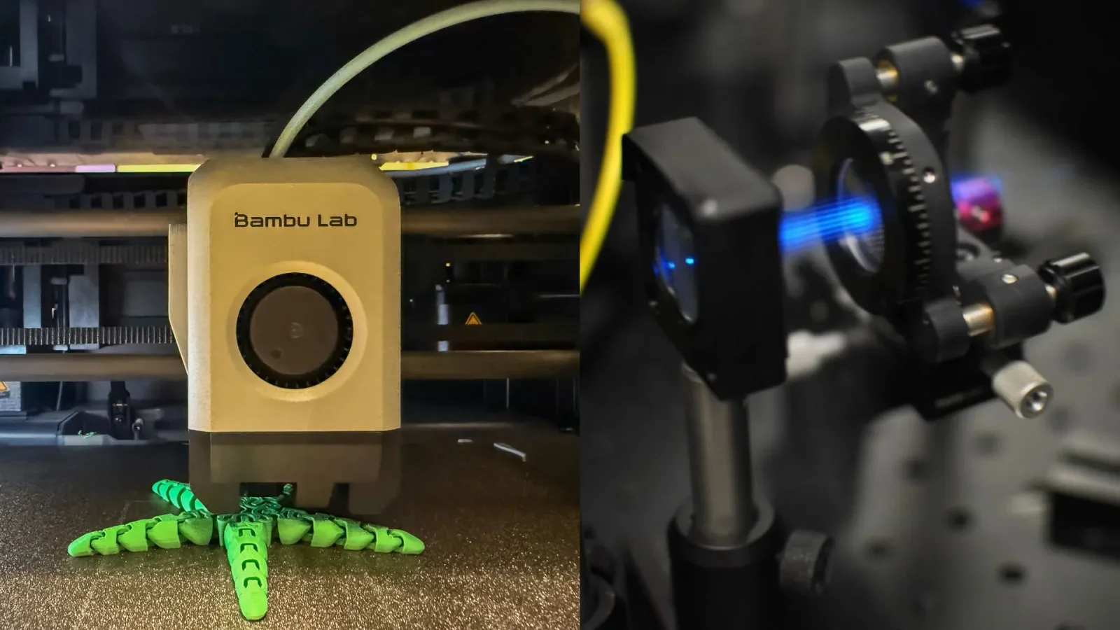
Researchers show that consumer-grade 3D printers, silicone molding, and UV-curable resin can create low-cost, multi-element lenslets for a multifocal structured illumination microscope, achieving about 150 nanometers resolution comparable to commercial systems and potentially democratizing access to high-performance imaging.
The 2025 Nikon Small World photomicrography competition, celebrating its 51st year, awarded first place to Zhang You for a detailed image of a rice weevil on rice, highlighting the blend of artistry and scientific rigor in microscopy.
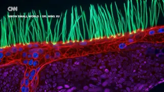
The article highlights the top microscopic photographs of 2025, showcasing stunning images of cellular structures, fungi, parasites, and insects captured through advanced microscopy techniques, emphasizing the beauty and complexity of the microscopic world.
Nikon's Small World contest showcases stunning microscopic images of tiny worlds, with this year's winners including a rice weevil, algae, and pollen, highlighting the beauty and complexity of small creatures and objects through advanced imaging techniques.
The 51st Nikon Small World Photomicrography Competition recognized 71 outstanding images captured through microscopy, with China's Zhang You winning first place for his detailed photo of a rice weevil on rice, highlighting the intricate world of insects and their ecological roles.
The article highlights award-winning microscopic images from the 2025 Nikon Small World competition, showcasing intricate details of tiny life forms like insects, cells, and microorganisms, emphasizing the importance of understanding and appreciating the complexity of microscopic worlds.
Scientists at Loughborough University have created the world's smallest violin, measuring just 35 by 13 microns, as a demonstration of their nanolithography capabilities, which could pave the way for advancements in computing and energy harvesting, though the violin is not playable.
Manu Prakash, a Stanford bioengineer, advocates for a philosophy called 'recreational biology,' which emphasizes curiosity and wonder in scientific exploration. He is renowned for creating low-cost scientific tools like the Foldscope, a paper microscope, and for studying both urgent health issues and the fascinating behaviors of microscopic organisms, blending practical applications with fundamental science to inspire broad curiosity and democratize access to scientific discovery.
Researchers at the Max Planck Institute have captured the first real-time video of ovulation, revealing the process in unprecedented detail. This breakthrough, achieved through advanced microscopy and live tissue culture techniques, allows scientists to observe the three key phases of ovulation, potentially revolutionizing infertility treatments. By understanding ovulation more precisely, this discovery could lead to improved diagnostics and treatments for conditions like polycystic ovary syndrome (PCOS), marking a significant advancement in reproductive medicine.
Scientists at the Max Planck Institute have successfully filmed the entire ovulation process in mice for the first time, using advanced microscopy techniques. This breakthrough allows researchers to observe the intricate dynamics of ovulation in unprecedented detail, potentially aiding in understanding conditions like polycystic ovary syndrome (PCOS) where ovulation is impaired. The study, published in Nature Cell Biology, marks a significant advancement in reproductive research.