
Mapping early β‑cell changes and immune interactions in type 1 diabetes progression
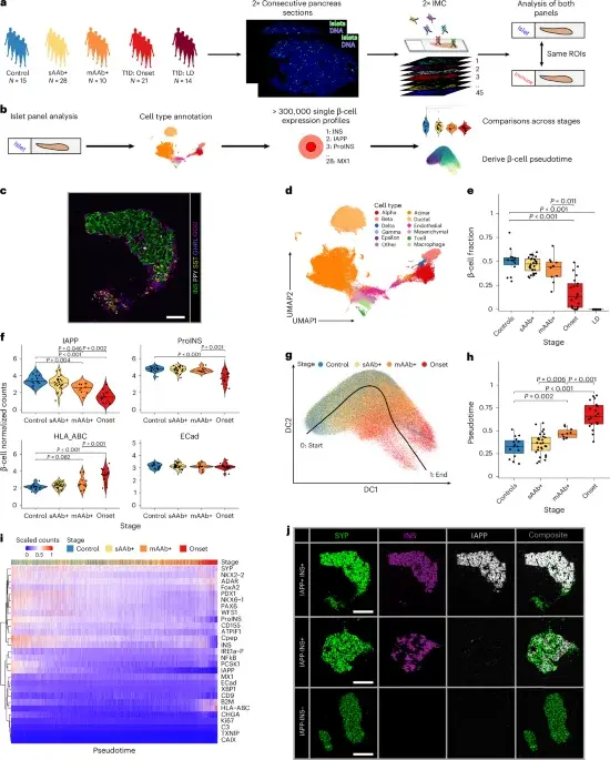
A large-scale imaging mass cytometry study of 88 human pancreata across the type 1 diabetes spectrum profiles 16 million cells to map β-cell states and the islet–immune interface. It finds β-cells show IAPP loss in preclinical stages and increased MHC-I at onset, with no universal rise in selected ER-stress markers. Insulitic islets display IFN signatures. Early disease features include pro‑inflammatory M1/M2‑like macrophages and exhausted‑like PD1+ TIM3+ T cells whose interactions occur at the islet edge and within islets, suggesting antigen presentation and regulatory dynamics. Age-related motifs reveal more TLS-like B/T cell structures and CD11c+ macrophages in younger donors. Pseudotime analysis suggests a single trajectory from healthy to insulitic β-cells, driven by macrophage–DC–exhausted T cell axis as disease progresses. The work provides a rich resource for understanding early T1D progression and identifying potential therapeutic targets and biomarkers.
- Imaging mass cytometry reveals functional and immunological changes during type 1 diabetes progression in human pancreata Nature
- UF Health researchers map early immune attack in Type 1 diabetes UF Health
- Unlocking diabetes’ secrets: Pediatric organ donors help map a path to a cure and prevention Vanderbilt Health News
- A spatial atlas of pancreatic remodelling in type 1 diabetes Nature
- Mapping histologic and functional maturation of human endocrine pancreas across early postnatal periods Nature
Want the full story? Read the original reporting
Read on Nature