
Shinbone Compression Triggers Brain Repair via Blood-Borne Signals
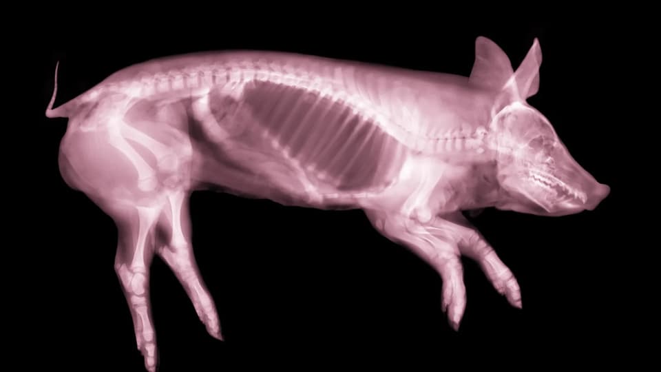
Researchers at Southern Medical University in China found that applying rhythmic pressure to the shinbone of mice and pigs with brain injuries significantly improved survival and neurological recovery. The study, published in Nature Neuroscience, demonstrates a bidirectional 'bone-brain axis' where mechanical stimulation of bone cells releases protective chemicals into the bloodstream that reduce inflammation and promote neuron repair in the brain.
Key points
- A team led by Zhiqing Cai used a technique called dynamic compressive tibial axial loading (DCTAL), applying pressure to the tibia 300 times per session at two pulses per second, five days a week.
- In mice, this treatment reduced neuron loss, lowered chronic inflammation, and improved performance in motor and spatial memory tests following both stroke and traumatic brain injury (TBI).
- The benefits depended on the PIEZO1 protein in osteocytes (bone cells); when this pressure-sensitive channel was genetically disabled, the protective effects disappeared.
- Serum from treated mice, containing factors like IL-1R2, APOL11a, HSP70, BDNF, and dopamine, replicated the brain-protective effects when injected into untreated injured mice.
- Pig experiments confirmed these findings in a larger animal model, showing improved survival and reduced brain damage, though the sample size was small (six per group) and limited to male animals.
Background
This research builds on the known observation that brain injuries can accelerate bone healing, suggesting a two-way communication system between the brain and skeleton. It follows earlier findings on skull immune hubs that link bone structures to brain health, reinforcing the concept of the skeleton as an active endocrine organ rather than passive scaffolding.
How outlets are covering it
ScienceAlert and ZME Science emphasize the novelty of the 'bone-brain axis' and the potential for future therapies that mimic bone signals without physical compression. Nautilus highlights the mechanical function of the PIEZO1 protein, describing its propeller-shaped structure that opens ion channels under pressure. While all sources agree on the efficacy in animal models, they collectively stress the limitations: the studies used only male animals, small pig sample sizes, and controlled lab injuries, meaning the approach is not yet ready for human application or real-world trauma scenarios.
Why it matters
This discovery opens a new therapeutic avenue for treating traumatic brain injuries and strokes by targeting the skeleton rather than the brain directly. If the specific chemical signals released by bone can be isolated or replicated, it could lead to non-invasive treatments that accelerate neurological recovery and reduce long-term damage from head injuries.
What to watch
Researchers must determine the optimal timing and duration for bone compression in humans, identify which specific blood factors are most critical for recovery, and conduct trials with female subjects and diverse injury types to ensure the findings translate to real-world clinical settings.
- Squeezing a Leg Bone Helped Animals Recover From Brain Injury ScienceAlert
- Tibial bone compression promotes recovery after brain injury through osteocyte PIEZO1 Nature
- How Gentle Tapping on the Shin Heals the Brain Nautilus | Science Connected
- Gentle rhythmic tapping on shinbone helps injured brains heal faster in animal models medicalxpress.com
- Compressing a Leg Bone Could Help Injured Brains Recover Faster zmescience.com
Want the full story? Read the original reporting
Read on ScienceAlert