
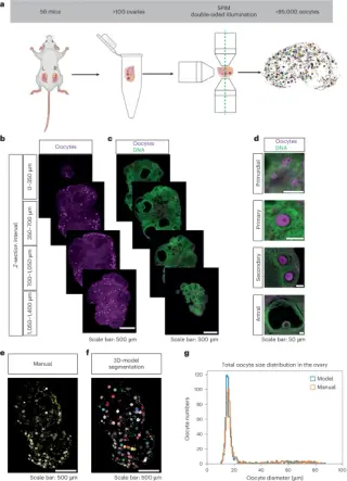
A 3D ovarian atlas reveals aging rules of the ovarian reserve
Using light-sheet microscopy and AI-driven segmentation to map more than 85,000 oocytes in whole mouse ovaries across the lifespan, the study finds a fixed fraction of oocytes activate despite overall decline, a lateral enrichment of follicles, and a bimodal size distribution suggesting a developmental bottleneck. A differential-equation model estimates primordial activation at ~2.5% per week with most oocyte loss occurring at the antral stage, revealing organ-wide regulatory dynamics of ovarian aging and a cross-species pipeline that also detects oocytes in human cortex.