
New Microfluidic Platform Maps 3D Genome Structure and Accessibility in Native Tissue
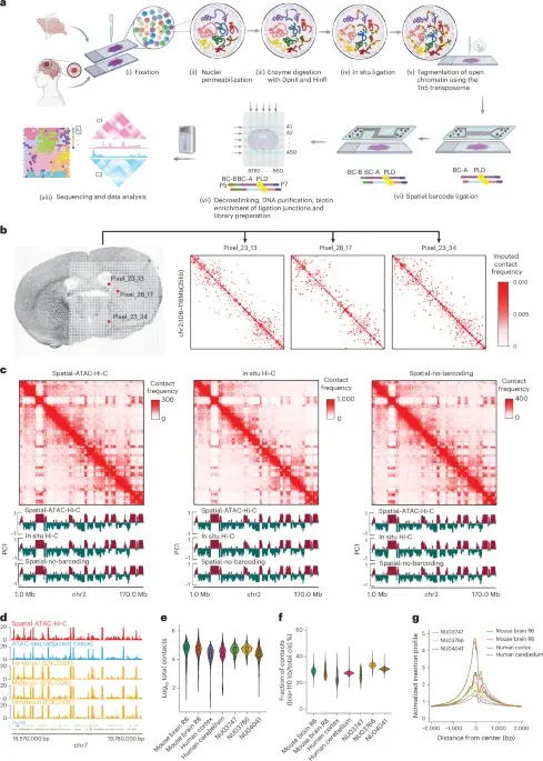
Researchers have developed Spatial-ATAC-Hi-C, a microfluidic technology that simultaneously profiles 3D genome organization and chromatin accessibility in intact tissue sections. Applied to mouse and human brains, the method reveals distinct regulatory programs in neuronal and non-neuronal cells. In glioblastoma and astrocytoma samples, it identifies spatially resolved copy number variations and structural variants, offering a new tool for studying tumor heterogeneity and gene regulation in native contexts.
Key points
- The new platform, Spatial-ATAC-Hi-C, uses barcoded oligonucleotides and microfluidic indexing to preserve spatial information while profiling 3D genome organization and chromatin accessibility.
- In adult mouse brains, the technology identified eight distinct clusters based on chromatin interactions and seven based on accessibility, which aligned with anatomical regions like the striatum and isocortex.
- The method successfully distinguished cell types, including excitatory neurons, inhibitory neurons, and non-neuronal cells, by identifying specific marker genes such as Satb2, Gad2, and Mpo.
- In human cerebellum samples, the platform confirmed cell-type-specific regulatory events, showing high chromatin interactions for granule neuron markers like ETV1 and NFIA.
- In cancer applications, the technology detected copy number variations and structural variants in glioblastoma and astrocytoma samples, revealing clonal heterogeneity and oncogenic events across tumor regions.
Background
This development builds on the decade-long evolution of spatial transcriptomics, which began with early 2016 work and has since expanded to include epigenomic and proteomic profiling. It complements recent findings on conserved euchromatin and diverse heterochromatin across eukaryotes, providing a new layer of spatial resolution for understanding how 3D genome organization influences gene regulation in health and disease.
Why it matters
By preserving native tissue architecture while simultaneously mapping 3D genome structure and chromatin accessibility, this technology allows researchers to study gene regulation in its natural spatial context. This is crucial for understanding complex biological processes like development and immune response, as well as for identifying clinically relevant oncogenic events and clonal heterogeneity in cancer, which are often missed by bulk or single-cell methods that lose spatial information.
What to watch
Researchers will likely apply Spatial-ATAC-Hi-C to other tissue types and disease models to further map spatial gene regulation. The ability to detect structural and copy number variations in native tissue may lead to new insights into tumor evolution and heterogeneity, potentially informing targeted therapies. Further validation and integration with other spatial omics platforms will be key to expanding its utility in clinical and basic research.
Want the full story? Read the original reporting
Read on Nature