
Cryo-Electron Microscopy Reveals Malaria Parasite’s Molecular Entry Mechanism
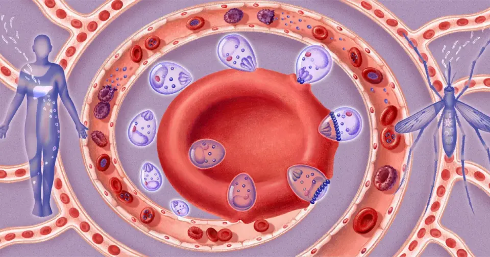
Researchers at Columbia University have used cryo-electron microscopy to capture the first detailed images of the 'moving junction,' a temporary molecular portal that allows Plasmodium parasites to invade human red blood cells. The study reveals that the parasite injects specific proteins that deform the host cell membrane through electrostatic forces, creating a gap that seals behind the invader. This structural insight offers a new target for antimalarial drugs, as blocking this entry point could prevent infection before the parasite establishes itself. While current treatments often rely on trial-and-error, this precise visualization of the invasion machinery may accelerate the development of targeted therapies for malaria and related parasitic diseases.
Key points
- Chi-Min Ho’s lab at Columbia University imaged the moving junction, a ring-shaped structure Plasmodium uses to enter red blood cells, using cryo-electron microscopy.
- The parasite injects three proteins that form a wedge beneath the cell membrane and a fourth protein connecting to the invader, using electrostatic forces to deform and reverse the membrane’s curvature.
- The invasion process takes less than one minute, after which the membrane seals behind the parasite, leaving no trace of the entry point.
- Blocking this invasion mechanism could serve as a new drug target, potentially preventing infection earlier than current treatments that target parasites already in the bloodstream.
- The moving junction machinery is shared by other apicomplexan parasites, including Toxoplasma gondii and Cyclospora, suggesting broad applications for drug discovery.
Background
Malaria, caused by Plasmodium parasites, remains a significant global health threat, killing approximately 600,000 people annually. While the basic life cycle of the parasite was identified in the late 19th century, the specific molecular mechanisms of cell invasion remained elusive due to the rapid and microscopic nature of the process. Previous attempts to image the invasion using electron microscopy in the 1970s only showed general membrane thickening, not the detailed protein structures involved. This new research builds on decades of incremental advances in microscopy and structural biology to finally visualize the dynamic process of membrane remodeling during invasion.
Why it matters
Understanding the precise structure of the moving junction provides a critical new target for antimalarial drug development. Current treatments often face rising resistance and lack mechanistic clarity, whereas blocking the invasion step could prevent infection entirely. Furthermore, since the same invasion machinery is used by other parasitic diseases like toxoplasmosis and cyclosporiasis, this knowledge could lead to broad-spectrum treatments for multiple parasitic infections, potentially saving lives and reducing the global burden of these diseases.
What to watch
Researchers plan to use similar cryo-electron microscopy techniques to analyze other protein complexes involved in malaria infection, such as adhesive proteins that help infected cells evade the spleen. Additionally, the team is developing AI-designed inhibitory proteins to block the moving junction, though these remain in early testing stages without animal model validation. The focus will shift to identifying stable drug candidates that can act within the brief 60-second window of the invasion process.
Want the full story? Read the original reporting
Read on Quanta Magazine