Tag

Cryo Electron Microscopy

All articles tagged with #cryo electron microscopy

science1 month ago

Megadalton Cytoplasmic Lattices Stockpile Embryo-Ready Proteins in Mouse Oocytes

Using cryo-electron microscopy and AI-based modeling, researchers reveal that mammalian oocyte cytoplasmic lattices (CPLs) are megadalton protein complexes formed from at least 13 components, including PADI6 and subcortical maternal complex members, incorporating unpolymerized tubulin and ubiquitination factors to stockpile essential proteins for early development; this explains how CPL disruption can lead to infertility and developmental defects.

science2 months ago

Cryo-EM unravels the filamentous blueprint of the oocyte cytoplasmic lattice

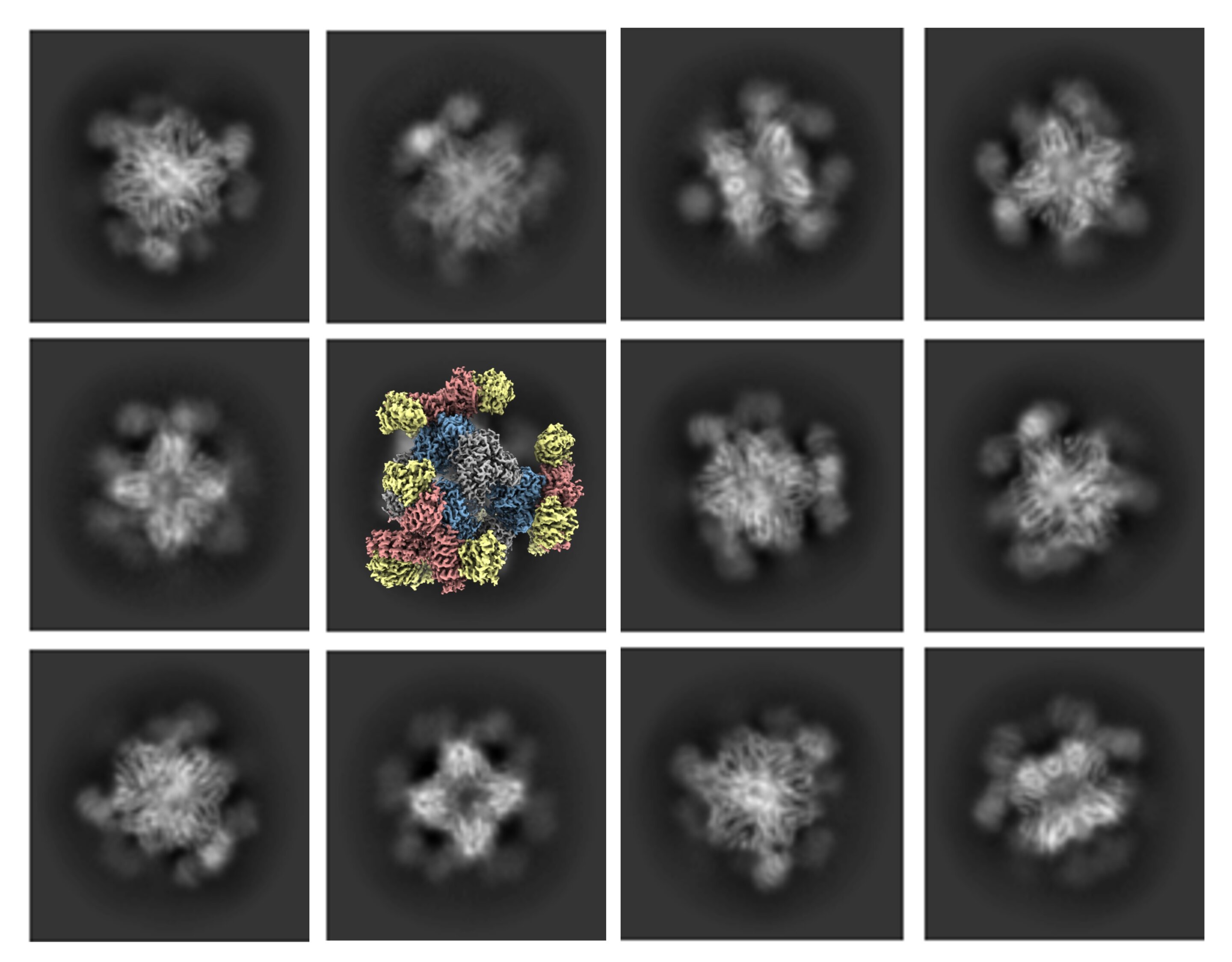
A mouse oocyte cytoplasmic lattice (CPL) structure was resolved by cryo-EM, revealing a repeating filament composed of U-shaped baskets and adapter rings built from 14 protein subunits; PADI6 anchors the baskets, while SCMC dimers connect adjacent units, outlining how CPL assembles and supporting its crucial roles in oocyte maturation, early embryogenesis, and potential female reproductive disorders.

science2 years ago

"Uncovering the Activation Mechanism of the Gabija Anti-Phage System"

The Gabija anti-phage system, a prokaryotic defense mechanism, consists of GajA and GajB components. Researchers have used cryo-electron microscopy to reveal the structures and activation mechanism of this system. They found that GajA, a rhombus-shaped tetramer, is activated for anti-phage defense when ATP is depleted by phages, leading to DNA cleavage. GajB, which docks on GajA, is then activated by the cleaved DNA, ultimately resulting in prokaryotic cell death.

Decoding the Intricate Molecular Structure of a Powerful Bacteriophage
science-and-technology2 years ago

Decoding the Intricate Molecular Structure of a Powerful Bacteriophage

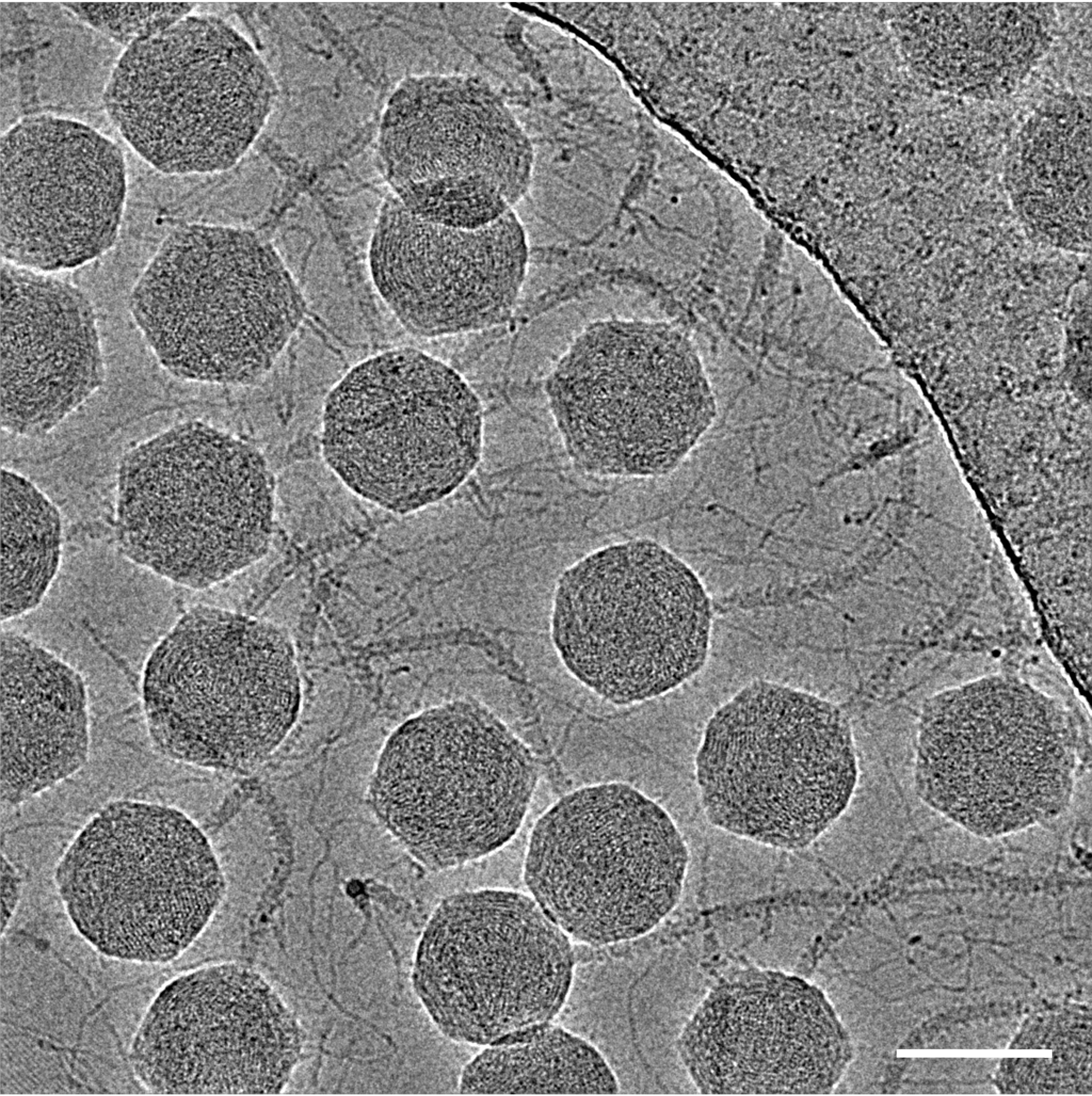
Scientists at the Okinawa Institute of Science and Technology have revealed the molecular structure of the tequintavirus, a type of bacteriophage that infects bacteria. Using cryo-electron microscopy, they obtained atomic models for all structural components of the virus, providing a detailed understanding of its organization at the atomic level. This research has implications for phage therapy, gene therapy, and the engineering of bacteriophages for specific purposes. The study also developed new methods for visualizing complex viruses, which could be applied to other viruses with similar shapes.

Unveiling the Link Between a Key Protein and Early-Onset Dementia
health2 years ago

Unveiling the Link Between a Key Protein and Early-Onset Dementia

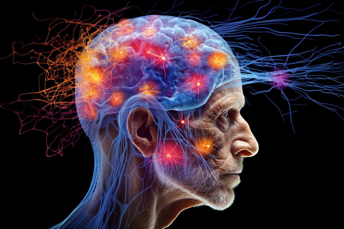
Researchers have identified the protein TAF15 as the key aggregate in certain cases of frontotemporal dementia, a type of neurodegenerative disease that affects the frontal and temporal lobes of the brain. Using advanced cryo-electron microscopy, scientists discovered TAF15 aggregates in brain samples, providing new insights into the molecular basis of the disease. This breakthrough could lead to targeted diagnostic tests and therapies for frontotemporal dementia, as well as potential treatments for motor neuron disease.

"Rapid Protein Mapping Achieved with Affordable Electron Microscope"
science-and-technology2 years ago

"Rapid Protein Mapping Achieved with Affordable Electron Microscope"

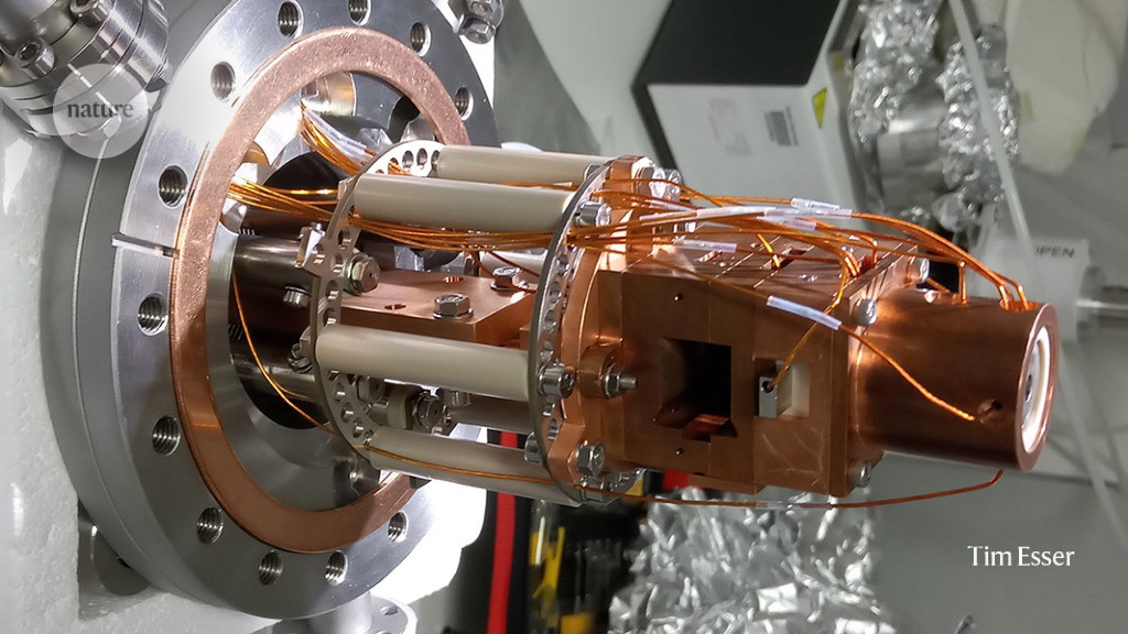
A newly designed electron microscope offers a cost-effective solution for mapping the 3D structures of cellular proteins, providing high-resolution images of biological molecules at a fraction of the cost of traditional models. The cryo-electron microscope allows for efficient analysis of cellular structures, such as the ribosome, and has the potential to revolutionize protein research.

Streamlining Structural Biology: Innovations in Soft-Landing Techniques
science-and-technology2 years ago

Streamlining Structural Biology: Innovations in Soft-Landing Techniques

Soft-landing mass spectrometry, a technique that gently lands intact proteins for analysis, shows promise in simplifying protein structure determination for cryo-electron microscopy (cryo-EM). By minimizing damage to proteins during the landing process, researchers have achieved near-atomic-resolution cryo-EM structures for proteins. This method could revolutionize protein sample preparation, allowing for the generation of high-resolution protein structures with greater precision and efficiency. However, further research is needed to optimize the technique and ensure that proteins retain their natural structure throughout the process. Soft-landing mass spectrometry also holds potential for single-molecule protein analysis and other structural analysis methods.

UCLA Team Advances Cryo-Electron Microscopy Technology
science-and-technology2 years ago

UCLA Team Advances Cryo-Electron Microscopy Technology

Scientists at UCLA have developed a solution to improve cryo-electron microscopy (cryo-EM) by enabling high-quality imaging of smaller protein molecules. They engineered a cube-shaped protein structure called a scaffold with tripod-like protrusions that hold the small proteins in place. The scaffold can be digitally removed during image processing, resulting in a composite 3D image of the small protein being analyzed. This advancement expands cryo-EM's imaging capabilities and has potential applications in drug development, allowing researchers to identify specific locations on proteins for therapeutic targeting. The technique was successfully tested on a protein involved in cancer treatments.

Unveiling the Origins of a Crucial Natural Molecule
science-and-technology2 years ago

Unveiling the Origins of a Crucial Natural Molecule

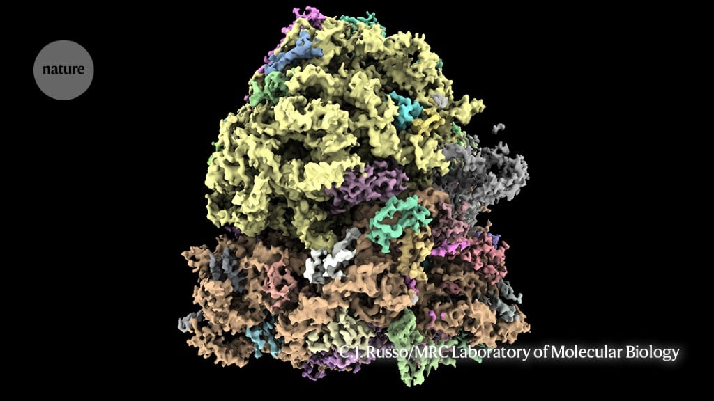
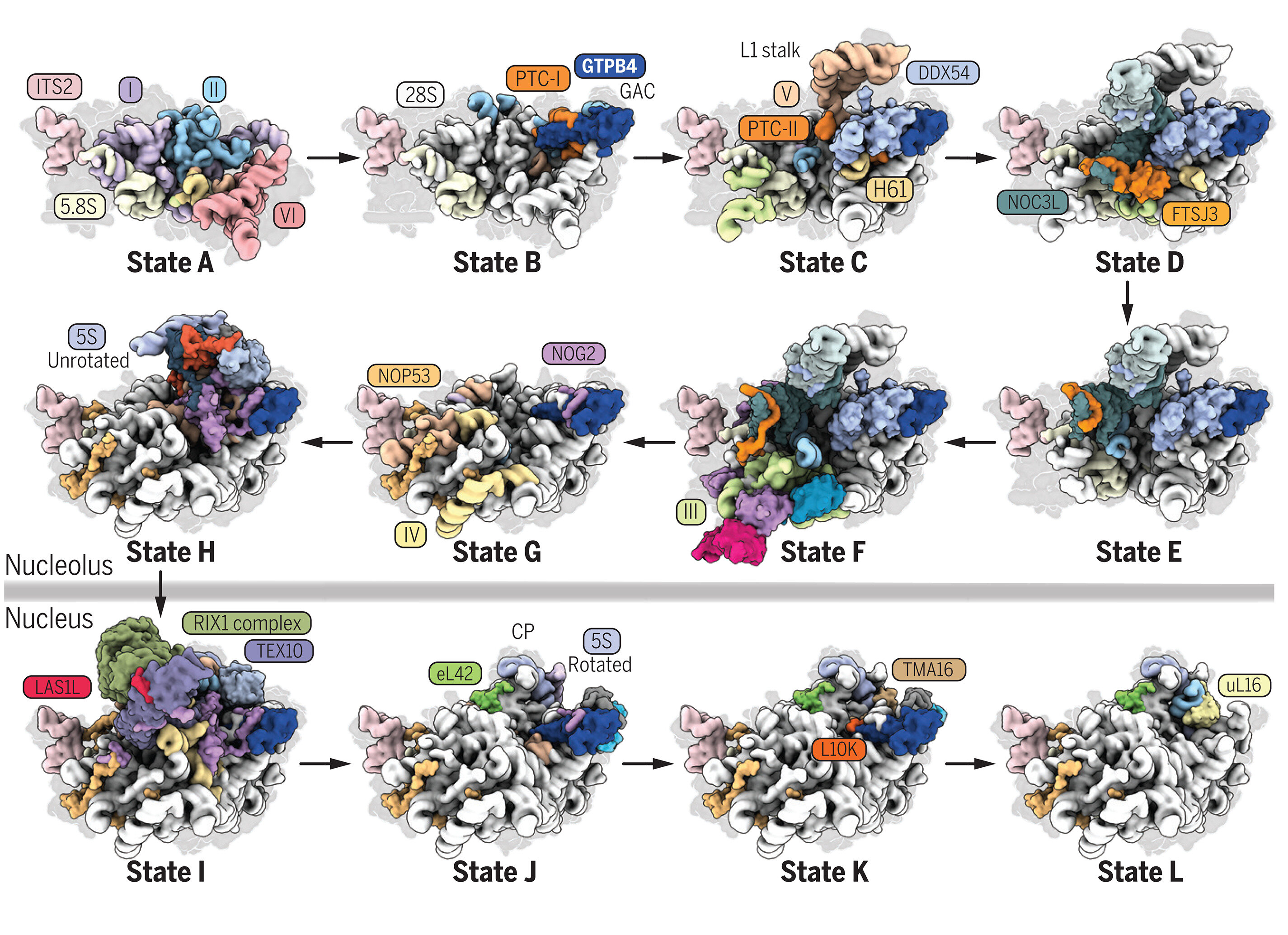
New high-resolution images obtained through cryo-electron microscopy have provided insights into the assembly of the large subunit of human ribosomes, shedding light on the formation of one of nature's most fundamental molecules. The study, which identified key steps and intermediates in the assembly process, could have implications for understanding cellular metabolism and diseases associated with ribosome mutations. The findings represent a significant advancement in our understanding of ribosome assembly and provide a foundation for further research in this field.

Unveiling the Formation of Nature's Fundamental Molecule
science-and-technology2 years ago

Unveiling the Formation of Nature's Fundamental Molecule

New high-resolution images of the large ribosomal subunit have provided insights into the assembly of one of nature's most fundamental molecules, ribosomes, in human cells. The study used cryo-electron microscopy to capture the formation and maturation of the human large ribosomal subunit (60S) and identified various proteins and enzymes that interact with RNA elements during the assembly process. The findings offer a near-complete picture of how the human large subunit assembles and could have implications for understanding cellular metabolism and diseases linked to ribosome mutations.