
Reproducible 3D human brain tissue model reveals mature microglia and Alzheimer's disease hallmarks
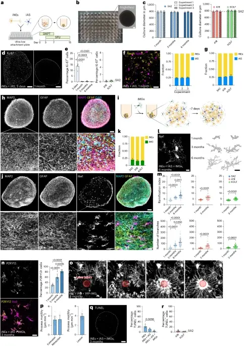
Researchers developed a reproducible iPSC-derived 3D cortical brain tissue model (3BTM) containing neurons, astrocytes and microglia that remains postmitotic and viable for months, forms brain-like ECM, and shows mature cell phenotypes. Microglia in 3BTMs display homeostatic features and in vitro–like in vivo responses. When engineered with APP mutations, 3BTMs recapitulate AD hallmarks—amyloid deposition, increased phospho-tau, and neuroinflammation—and exhibit disease-associated microglial signatures. Adding exogenous Aβ42 forms plaque-like structures, while treatment with aducanumab reduces Aβ load and enhances microglial activity, validating a translational drug-testing platform. Compared with 2D cultures, 3BTMs show greater maturation and network functionality across multiple iPSC lines, enabling scalable studies of brain physiology, disease mechanisms, and therapeutic responses.
- A reproducible three-dimensional model of human brain tissue to investigate physiological and disease-associated microglia phenotypes Nature
- A new dimension in brain research AIP.ORG
- 3D Human Brain Tissue Model Replicates Alzheimer’s Pathology neurosciencenews.com
- Reproducible 3D Human Brain Tissue Model Studies Healthy and Disease-Linked Microglia Phenotypes Bioengineer.org
- New 3D brain tissue model accelerates Alzheimer's drug development News-Medical
Want the full story? Read the original reporting
Read on Nature