
New Deep Learning Method VIMA Identifies Disease-Specific Spatial Patterns in Tissues
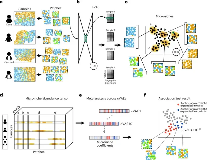
Researchers have introduced VIMA, a new computational method that uses deep learning to analyze spatial molecular data in tissues. By creating numerical 'fingerprints' for small tissue patches, VIMA identifies disease-associated structures without requiring manual cell segmentation or discrete clustering. Tested on rheumatoid arthritis, ulcerative colitis, and dementia datasets, the tool successfully recapitulated known biology and revealed new spatial features, outperforming existing methods in detecting case-control differences.
Key points
- VIMA employs an ensemble of variational autoencoders to generate numeric fingerprints for tissue patches, avoiding restrictive assumptions about discrete cell types or niches.
- The method defines overlapping 'microniches' to quantify abundance and performs statistical tests to correlate these with case-control status, offering high power even with small sample sizes.
- In rheumatoid arthritis, VIMA distinguished stromal from immune subtypes using only seven markers, revealing spatial heterogeneity within individual samples that single-cell analysis missed.
- For ulcerative colitis, the tool identified that TNF inhibition remodels colonic architecture by reducing lymphoid aggregates and increasing mesenchymal activity, a signal undetected by other benchmarked methods.
- VIMA requires no pretraining, has a favorable runtime of approximately 2 to 8 hours per dataset on a single GPU, and produces calibrated P-values for hypothesis testing.
Background
This development follows recent advancements in spatial molecular data analysis, where researchers are increasingly moving beyond discrete cell-type abundances to capture complex tissue microenvironments. While previous methods relied on manual scoring or hard clustering, which can obscure subtle signals, VIMA represents a shift toward flexible, data-driven representations. This aligns with broader trends in biomedical research to leverage high-resolution spatial modalities, such as CODEX and MERFISH, for more precise disease characterization and precision medicine applications.
Why it matters
VIMA addresses a critical bottleneck in spatial biology: the inability of current methods to systematically detect disease-associated spatial structures without restrictive preprocessing. By eliminating the need for cell segmentation and discrete clustering, it enables researchers to analyze complex, high-dimensional spatial data with greater precision. This could accelerate the discovery of new disease mechanisms and biomarkers, particularly in conditions where spatial organization of cells is crucial, such as cancer and autoimmune diseases.
What to watch
The developers have released open-source software for VIMA, allowing the broader research community to apply the method to new datasets and technologies. Future work will likely focus on expanding the tool to additional spatial modalities and larger cohorts, potentially integrating it with other omics data to provide a more comprehensive view of tissue biology. The method's ability to handle modest sample sizes suggests it could be widely adopted in clinical and research settings where large cohorts are not always feasible.
Want the full story? Read the original reporting
Read on Nature