Tag

Spatial Transcriptomics

All articles tagged with #spatial transcriptomics

Spatial atlas reveals how human organs begin to form after gastrulation
science3 days ago

Spatial atlas reveals how human organs begin to form after gastrulation

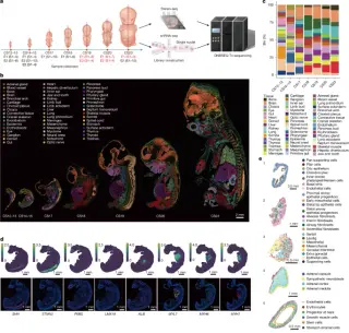
Pan et al. create a comprehensive spatiotemporal atlas of gene expression in 13 human embryos from Carnegie stage 12 to 23 using Stereo-seq and snRNA-seq, mapping 50 organs and 198 substructures, revealing context-specific regulatory networks and tissue identities, uncovering new gene functions in cardiac and brain development, and providing a public resource to visualize the embryonic transcriptional landscape and allelic expression dynamics across development.

TNBC’s heterogeneity mapped: four cancer archetypes and eight tissue ecotypes forecast chemo response
health17 days ago

TNBC’s heterogeneity mapped: four cancer archetypes and eight tissue ecotypes forecast chemo response

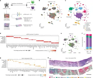
A large single‑cell and spatial transcriptomics study of pretreatment TNBC identifies four cancer-cell archetypes and eight ecotypes defined by cancer cells and the tumor microenvironment, reveals macrophage and interferon‑related programs linked to better neoadjuvant chemotherapy response, and provides cell-state– and gene-based classifiers (including a 13‑gene panel) to predict pathological complete response, offering new insight into TNBC heterogeneity and potential immune targets such as LAG3 and HAVCR2.

Scar-Driven Fibrosis Emerges as Target for Treatment-Resistant Rheumatoid Arthritis
health27 days ago

Scar-Driven Fibrosis Emerges as Target for Treatment-Resistant Rheumatoid Arthritis

A Nature Immunology study using spatial transcriptomics reveals that persistent scar formation (fibrosis) and disrupted fibroblast–endothelial signaling in RA joints may drive treatment resistance even when inflammation is controlled, pointing to a new fibrosis-focused therapeutic target for patients who don’t respond to standard therapies.

B cell–driven Diabetic Kidney Disease uncovered by a high-resolution spatial atlas
science1 month ago

B cell–driven Diabetic Kidney Disease uncovered by a high-resolution spatial atlas

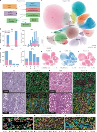
Researchers built a cross-platform spatial atlas of diabetic kidney disease using CosMx and Xenium, integrated with snRNA-seq to define 11 tissue niches and injury microenvironments. They identify a B cell–predominant immune microenvironment that defines a DKD subset with faster kidney function decline, and develop tissue biomarkers plus a plasma protein panel that improve risk prediction beyond clinical models; findings validated in UK Biobank and supported by plasma proteomics, highlighting biomarker-guided B cell–targeted DKD trials.

Live-donor atlas maps healthy liver zonation and early steatosis dynamics
science1 month ago

Live-donor atlas maps healthy liver zonation and early steatosis dynamics

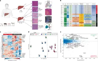
Researchers built a high‑resolution spatial atlas of the healthy human liver from live donors, integrating Visium, MERFISH and single-nucleus RNA‑seq to map hepatocyte zonation along the porto–central axis and compare with adjacent diseased tissue. They validate distinct pericentral and periportal programs, reveal early steatosis signatures such as reduced nuclear-encoded mitochondrial genes with compensatory mitochondria-encoded transcripts, and extend analyses to other mammals. The atlas provides a healthy-liver baseline to advance biology and disease research.

Skin’s cellular map reveals TNF-driven immune–fibroblast networks in SALT-like neighborhoods
science2 months ago

Skin’s cellular map reveals TNF-driven immune–fibroblast networks in SALT-like neighborhoods

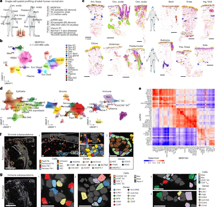
Researchers generated an organ-wide, MERFISH-based single-cell spatial atlas of about 1.2 million cells from normal adult human skin, spanning 15 body sites and 22 donors, resolving 45 cell types and 10 multicellular neighborhoods. They highlight a perivascular neighborhood reminiscent of skin-associated lymphoid tissue (SALT) where TNF coordinates immune–fibroblast crosstalk, with CCL19+ fibroblasts proximal to vessels playing a central role. By integrating MERFISH with scRNA-seq and Visium data, they map site-specific cellular compositions, neighborhood dynamics, and ligand–receptor signaling, and show pan-disease immune alterations in the perivascular niche across skin conditions. An interactive web tool is provided to explore these data, underscoring spatial organization as a key driver of skin biology and disease.

Developmental cell states drive heterogeneity in supratentorial ependymomas
science2 months ago

Developmental cell states drive heterogeneity in supratentorial ependymomas

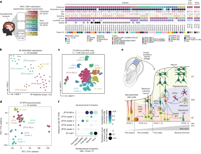
A multidimensional study integrating single-cell and spatial transcriptomics with live-cell imaging reveals two progenitor-like states—neuroepithelial-like and embryonic-like—and diverse spatial architectures in supratentorial ependymomas. Brain-resident cells can shift tumor cells toward neuronal-like, migratory phenotypes, highlighting developmental origins of heterogeneity and suggesting potential therapeutic angles.

Mapping the Developing Human Heart to Understand Congenital Defects
science7 months ago

Mapping the Developing Human Heart to Understand Congenital Defects

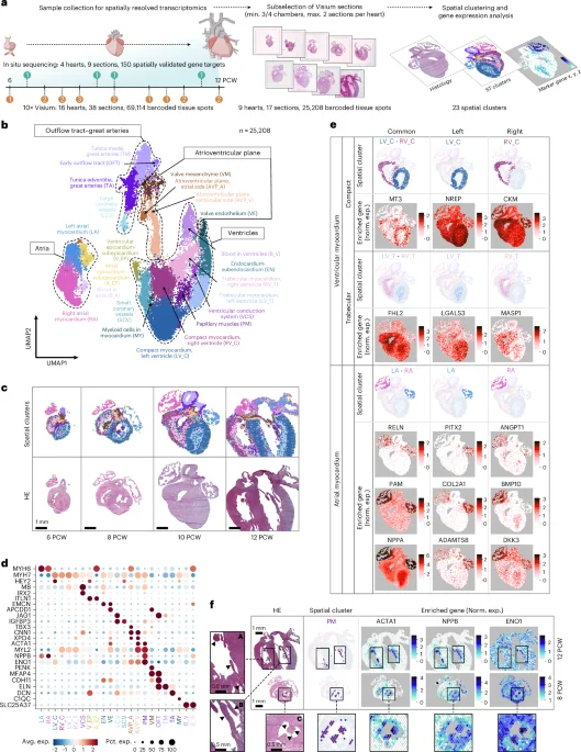
This study provides a detailed spatiotemporal map of gene expression and cellular dynamics during early human heart development, analyzing over 69,000 tissue spots and nearly 77,000 cells across 36 hearts, revealing diverse cell types, specialized conduction system components, and the formation of cardiac innervation and valves, thereby advancing understanding of human cardiogenesis.

Unveiling the Intricate Development of Human Limbs: From Embryonic Atlas to Nature's Chiseling
science-and-technology2 years ago

Unveiling the Intricate Development of Human Limbs: From Embryonic Atlas to Nature's Chiseling

Researchers have created a comprehensive atlas of human embryonic limb development using single-cell transcriptomic RNA sequencing and spatial transcriptomic sequencing. The study identified 67 distinct cell clusters and mapped their spatial distribution across four timepoints during the first trimester of development. The research sheds light on the cellular heterogeneity, patterning events, and gene expression patterns associated with limb malformations. The findings also highlight the similarities between human limb development and that of model organisms like mice. This atlas provides valuable insights into the complex processes involved in limb development and can serve as a reference for studying genetic variations and developmental disorders.

Unraveling the Inflammatory Puzzle: How Obesity and Immune Cells Interact in Fat Tissue
health2 years ago

Unraveling the Inflammatory Puzzle: How Obesity and Immune Cells Interact in Fat Tissue

A study conducted by researchers at the University of Michigan has revealed new insights into the complex inflammatory response that occurs within fat tissue in obesity. Using single cell analysis and spatial transcriptomics, the study identified previously unrecognized immune cell types and interactions within adipose tissue. The researchers discovered different subtypes of macrophages, with some exhibiting pro-inflammatory genes and others showing low pro-inflammatory gene expression. The findings suggest that the body may attempt to counter inflammation by promoting lipid-associated macrophages. Further research will focus on understanding the signaling processes and proteins associated with the development of these macrophages and metabolic disorders.