Novo Nordisk signaled a bigger M&A push and ambitions for more than five obesity blockbusters by 2030, a plan that led to a share drop for Novo while lifting smaller obesity-focused names VKTX, GPCR and ALT on expectations of deal activity and partnerships.
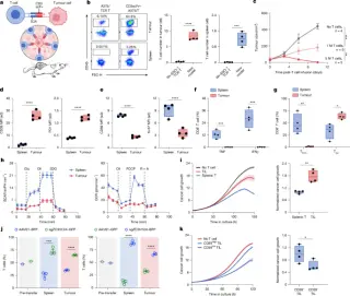
Nature reports a scalable in vivo platform for genome-wide CRISPR screens in human T cells within solid tumors. Two screens found that GPCR signaling via the P2RY8–Gα13 axis limits T cell tumor infiltration, while GNAS (Gαs) drives intratumoral T cell dysfunction. Knocking out GNAS enhances T cell fitness and antitumor efficacy across multiple CAR/TCR models, and combining P2RY8 and GNAS edits yields even better tumor control, enabling systematic genetic improvements for solid-tumor T cell therapies.
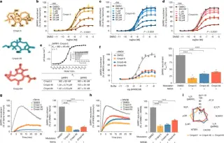
Nature reports the discovery of the first small-molecule inhibitors that directly bind and inhibit β-arrestins, blocking their engagement with agonist-activated GPCRs and downstream signaling while sparing G protein coupling. Using differential scanning fluorimetry, three modulators (Cmpd-5/oridonin, Cmpd-46, Cmpd-64) were characterized across biophysical and cellular assays, showing dose-dependent inhibition of β-arrestin recruitment and receptor desensitization/internalization. Cryo-EM reveals Cmpd-5 binding to a central crest (the MCL site) on β-arrestin1, stabilizing an inactive-like conformation incompatible with receptor engagement. Complementary ITC, MD simulations, docking, and mutagenesis validate this allosteric pocket as a drug-design target. Across GPCR panels, T cell migration, and cardiomyocyte assays, these modulators alter β-arrestin signaling and effector interactions without suppressing Gi or Gs activity, suggesting a path toward pathway-specific GPCR therapeutics. Data include PDB/EMDB structures for βarr1–Cmpd-5 and related states, and the work outlines a mechanistic framework for transducer-targeted GPCR drugs.
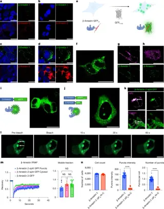
New research shows beta-arrestins 1 and 2 form endogenous condensates near activated GPCRs via liquid–liquid phase separation, a process regulated by IP6-induced oligomerization and the arrestin intrinsically disordered region. These condensates influence GPCR desensitization, internalization, and downstream signaling, with arrestin orientation and specific mutations (IP6 sites and IDR) altering function, suggesting LLPS compartments are important regulators of GPCR signaling and could inform drug development.
Nature reports computational de novo design of miniproteins that bind G protein-coupled receptors (GPCRs) with high affinity, enabling both agonists for itch/pain receptors and antagonists for cancer, metabolic disorders, and migraine. Cryo-EM structures of five receptor–miniprotein complexes closely match the design models, validating the approach, and a designed chemokine receptor antagonist mobilizes hematopoietic stem and progenitor cells in vivo with fewer adverse effects than a clinically used drug.
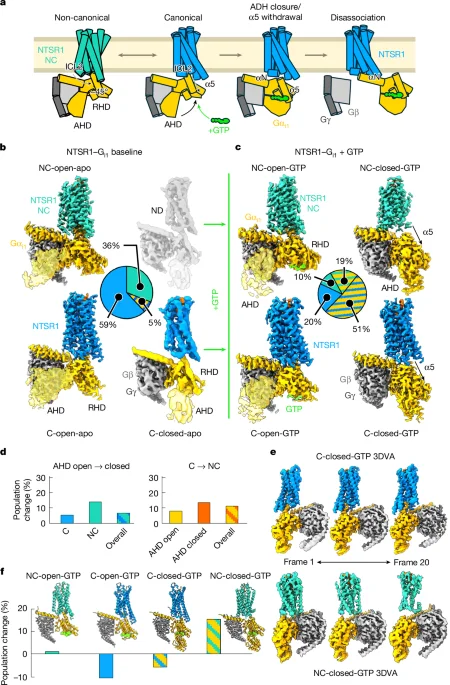
Time-resolved cryo-EM maps reveal multiple GTP-bound intermediate states for the NTSR1–Gi1 and NTSR1–G11 complexes, showing that receptor intracellular loops ICL2 and ICL3, along with G protein regions, shape subtype selectivity beyond nucleotide-free structures. Gi1 forms more stable intermediates and dissociates slower than G11, explaining differential signaling, and swapping ICL2/ICL3 between NTSR1 and MOR disrupts these intermediates and signaling, highlighting a dynamic, intermediate-state mechanism underlying GPCR–G protein coupling and selectivity.
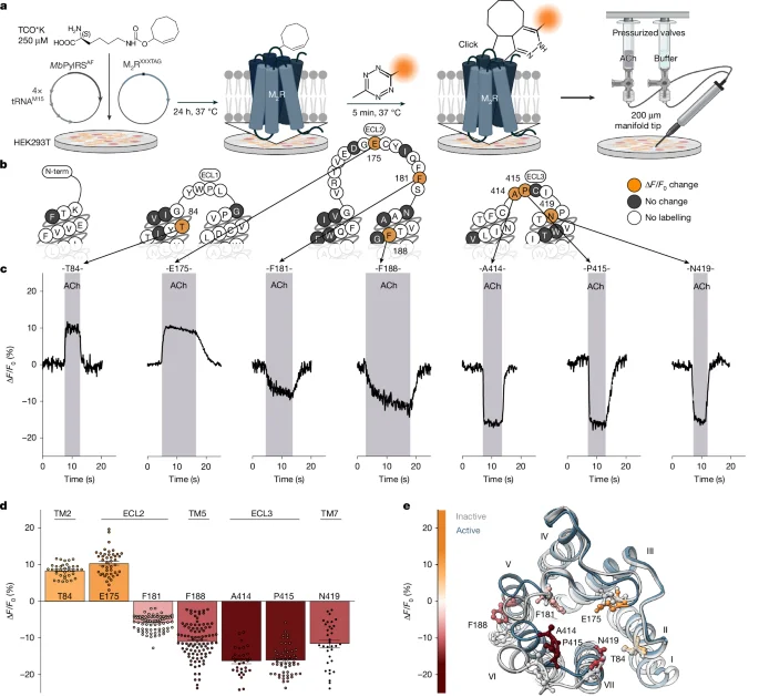
Researchers built extracellular, single-residue GPCR biosensors on the M2 muscarinic receptor using noncanonical amino acids and fast click labeling to watch real-time, ligand-induced conformational changes in live cells. They show agonists stabilize at least two distinct M2R–G-protein signaling complexes (C1 and C2) that form via ligand-specific activation trajectories, with the complex balance and trajectory dictating G-protein subtype selectivity and signaling strength. Overexpressing a nucleotide‑free Gα mutant shifts equilibria to reveal different complexes, while PTX and TRUPATH assays map GDP-bound, low-efficacy and GDP-free, high-efficacy states and their G-protein preferences. Kinetic analysis links on-rates (0.2–5 s formation) to activation trajectories, suggesting ligand-specific pathways underlie GPCR signaling diversity and offering new angles for drug discovery.
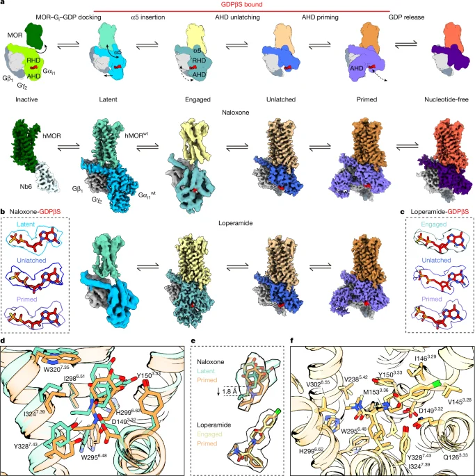
The study reveals how different ligands stabilize distinct conformations of the μ-opioid receptor, affecting GDP release rates and G protein activation, with structural snapshots and simulations supporting a model where ligand efficacy influences receptor conformational states and signaling outcomes.
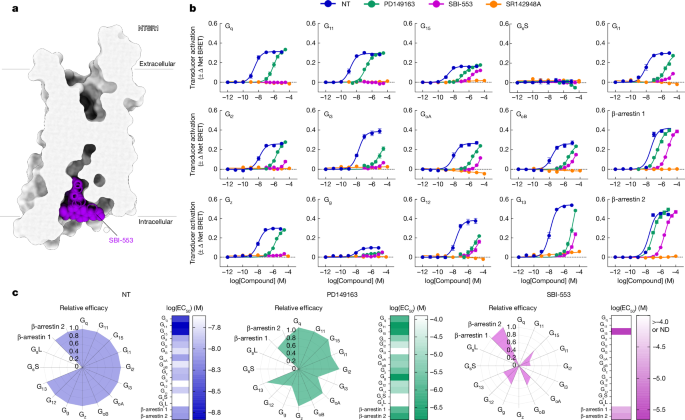
The article discusses the design of allosteric modulators targeting GPCRs, specifically NTSR1, to alter G protein subtype selectivity and biased signalling, demonstrating that minor structural modifications can predictably change receptor coupling profiles and in vivo effects, paving the way for more targeted therapeutics.
Researchers have identified a molecular complex, Alex3/Gαq, crucial for the transport of mitochondria within neurons, which is essential for neurotransmission and neuronal functions. This finding offers potential therapeutic avenues against neurodegenerative diseases, neuromuscular disorders, and certain cancers by targeting the mitochondrial transport mechanism. The study underscores the complex’s role in maintaining neuronal health and viability, suggesting that manipulating this system could offer innovative treatments for a range of diseases linked to mitochondrial dysfunction.
Researchers have discovered a new way to activate G-protein coupled receptors (GPCRs) from inside cells, using a molecule called PCO371 that interacts with the intracellular region of a specific GPCR. This method could help develop drugs with fewer or no side effects for conditions such as obesity, pain, osteoporosis, and neurological disorders. The specificity of PCO371's binding and receptor activation mode makes it a suitable candidate for potential small-molecule-based drugs for class B1 GPCRs, which currently lack oral administrative drug ligands.