Tag

Amygdala

All articles tagged with #amygdala

Why traumatic memories come in fragments and feel like the present moment
health5 days ago

Why traumatic memories come in fragments and feel like the present moment

Traumatic memories are not stored or recalled like ordinary memories; during trauma, the amygdala stays highly active while the hippocampus is overwhelmed, causing memories to break into sensory fragments and to be relived as if the threat is happening now. This non-linear recall can look inconsistent or even be misread as lying, especially by lawyers or clinicians. A trauma‑informed approach—empathy, careful language, and awareness that memory can be fragmented—improves support in healthcare, legal settings, and everyday relationships and can aid survivors’ healing.

Big Amygdala, Subtle Bias: Brain Structure and Our Reactions to the Unknown
science10 days ago

Big Amygdala, Subtle Bias: Brain Structure and Our Reactions to the Unknown

Neuroimaging links larger amygdala and stronger responses to unfamiliar faces with conservative leanings and threat sensitivity, but large replication studies show the effect is small and not diagnostic; the amygdala may function as a significance detector rather than a fear center, habituation reduces responses to known faces, and cross-group contact generally reduces prejudice, suggesting biology partly influences discomfort with difference but isn’t destiny.

Migraines May Accelerate Brain Aging in Memory and Emotion Regions
science21 days ago

Migraines May Accelerate Brain Aging in Memory and Emotion Regions

New research suggests migraines are linked to accelerated brain aging: migraine patients show a global brain-age gap of about 4.24 years and 66 regions with higher aging, concentrated in memory- and emotion-related areas (prefrontal/frontal/parietal/temporal cortices and the amygdala). The findings come from cross-sectional MRI analysis and do not prove causation; results may not generalize to all migraine sufferers or reflect cognitive performance without further testing.

Brain circuit links social memory to fear, triggering aversion in mice
science-and-health1 month ago

Brain circuit links social memory to fear, triggering aversion in mice

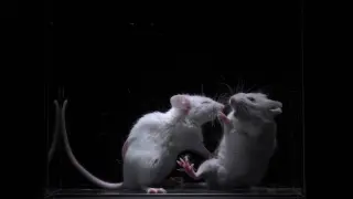
University of Tokyo researchers identify a hippocampus–amygdala circuit that binds the memory of a familiar mouse to fear, causing avoidance after aggressive encounters; using optogenetics they could strengthen or erase this aversion, with the nucleus accumbens helping translate memory and fear into avoidance. While demonstrated in mice, the findings offer clues about how social memory and negative emotions interact, with potential relevance for anxiety and depression in humans.

Timing of Childhood Trauma Shapes Distinct Brain Signatures in Adulthood
neuroscience1 month ago

Timing of Childhood Trauma Shapes Distinct Brain Signatures in Adulthood

A study of 635 adults using fMRI found that the age at which abuse occurs matters for later emotion processing: abuse before age 13 is linked to heightened hippocampal activity during rapid, non-conscious emotion processing, while abuse in adolescence (13–18) is linked to heightened amygdala activity during conscious emotion processing. These patterns persisted across various mental health diagnoses and healthy controls, suggesting the timing of adversity leaves lasting neural marks and could guide targeted interventions, though retrospective reporting and cross-sectional design limit conclusions.

Timing of Childhood Trauma Rewires Brain–Immune Fear Circuits
science2 months ago

Timing of Childhood Trauma Rewires Brain–Immune Fear Circuits

A 128-participant fMRI study from Guangzhou University links the type and timing of childhood maltreatment to lasting neuro-immune changes: early childhood abuse (ages 1–11) modulates amygdala–IL-8 coupling during fear learning, late adolescence neglect (ages 12–18) reshapes vmPFC–IL-8 connections, and early neglect alters amygdala–vmPFC and hippocampus–vmPFC connectivity with IL-17; the findings support a maturation-matched vulnerability and point to therapies that target both fear circuits and systemic inflammation.

Targeted amygdala circuit tweak reverses anxiety in mice
science2 months ago

Targeted amygdala circuit tweak reverses anxiety in mice

A study in iScience links imbalanced activity of a Grik4/GluK4-rich neuron group in the basolateral amygdala to anxiety-like and social withdrawal behaviors in mice. Restoring this balance to improve signaling with centrolateral inhibitory neurons reversed anxiety and social deficits in both a genetic model and wild-type mice with high anxiety, though some memory issues persisted, hinting at broader circuit targets for affective disorders.

Long COVID Not Driven by Widespread Brain Inflammation; Emotions Centers Linked to Symptom Severity
health2 months ago

Long COVID Not Driven by Widespread Brain Inflammation; Emotions Centers Linked to Symptom Severity

Finnish researchers used TSPO-PET and MRI to scan 14 long COVID patients, 11 healthy controls, and 13 MS patients, finding no evidence of widespread brain inflammation in long COVID and suggesting inflammatory activity may be higher earlier after infection and fade over time. Notably, more severe symptoms correlated with increased activity in emotion-regulation regions like the amygdala and hippocampus, pointing to emotional processing rather than ongoing neuroinflammation as a potential driver and hinting that treatments could focus on stress management and emotional regulation rather than solely anti-inflammatory approaches. The study is published in the Journal of Neurology (2026).

Night Shifts Subtly Shrink Brain Regions, But Reversals Occur After Stopping
science2 months ago

Night Shifts Subtly Shrink Brain Regions, But Reversals Occur After Stopping

A large UK Biobank MRI study found that among 2,122 shift workers, there is a modest, symmetrical loss of brain volume in the right thalamus and left amygdala (areas tied to sleep regulation, emotion, and memory) with associated white‑matter changes. The negative association with cognitive performance is small, and the researchers caution interpretation. Importantly, ceasing shift work was linked to partial recovery of brain volume within about 2.5 years, suggesting a potential reversible window, though the study focused on older adults and applicability to younger workers remains uncertain.

Long COVID Symptoms Linked to Mood-Processing Brain Activity, Not Widespread Inflammation
health-and-medicine2 months ago

Long COVID Symptoms Linked to Mood-Processing Brain Activity, Not Widespread Inflammation

A University of Turku study using PET and MRI found no evidence of widespread brain inflammation in long COVID patients compared with healthy controls. Inflammation may be higher earlier after infection and fade over time. More severe symptoms correlated with increased activity in emotion-related regions (hippocampus and amygdala), suggesting treatments focusing on stress management and emotional regulation rather than solely anti-inflammatory approaches.

Shift Work Linked to Subtle Shrinkage in Amygdala and Thalamus, Reversible Upon Stopping
neuroscience2 months ago

Shift Work Linked to Subtle Shrinkage in Amygdala and Thalamus, Reversible Upon Stopping

An analysis of UK Biobank data found that people who work shifts show a small but detectable volume loss in the left amygdala and right thalamus compared with non-shift workers, with greater loss at higher shift-work frequency. In participants who ceased shift work, the brain-volume decline halted within about 2.4 years and may show slight recovery; additional microstructural changes and lower scores on memory, processing speed, and fluid intelligence were observed. The authors caution that causality cannot be established and the effects are small, noting the UK Biobank sample is healthier and less diverse than the general population.

Psilocybin’s 5-HT2A activation linked to lasting brain plasticity in mice
neuroscience5 months ago

Psilocybin’s 5-HT2A activation linked to lasting brain plasticity in mice

A mouse study shows psilocybin dose-dependently activates the brain’s 5-HT2A receptors in the prefrontal cortex, with an inverted-U relationship for acute behaviors. The following day, moderate doses reduced anxiety-like exploration and higher doses decreased depression-like immobility, coinciding with changes in microtubule dynamics and increased synaptic plasticity proteins—primarily in the prefrontal cortex, not the amygdala—suggesting a neural mechanism for lasting antidepressant effects, though results in animals may not directly translate to humans.