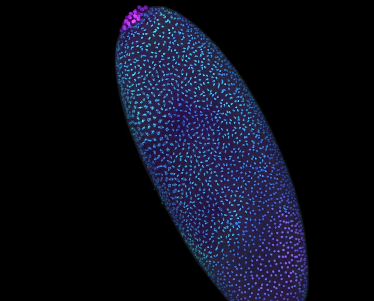
A study of the sea anemone Nematostella vectensis shows it uses a BMP-Chordin shuttling system to shape its embryo, revealing an ancient, shared mechanism for organizing body plans that predates the split between cnidarians and bilaterians.
Researchers using the Pico-C method show that a modular 3D genome scaffold forms in Drosophila embryos before zygotic genome activation, suggesting pre‑existing DNA organization guides timely gene activation and development; disrupting this architecture can trigger immune-like responses, highlighting its importance for human health as well as early-life biology.
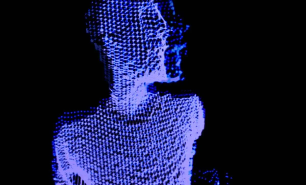
Researchers from the Brugués group at TU Dresden report in Nature a new mechanism for early embryonic cell division in yolk-rich cells: a mechanical ratchet that drives division without a fully closed actin contractile ring. By showing microtubule asters stiffen the cytoplasm during interphase and the cytoplasm becomes more fluid in M-phase, they find the actin band can ingress across multiple cell cycles, anchored by microtubules and re-stabilized when the cytoplasm stiffens again. This challenges textbook models and may apply broadly to yolk-rich embryos across species.
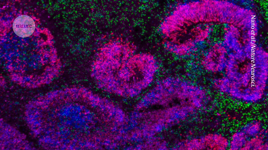
Scientists have created the most detailed maps of how human and mouse brains develop from stem cells into neurons, providing new insights into brain differentiation and potential implications for neurological conditions, as part of the BRAIN Initiative.
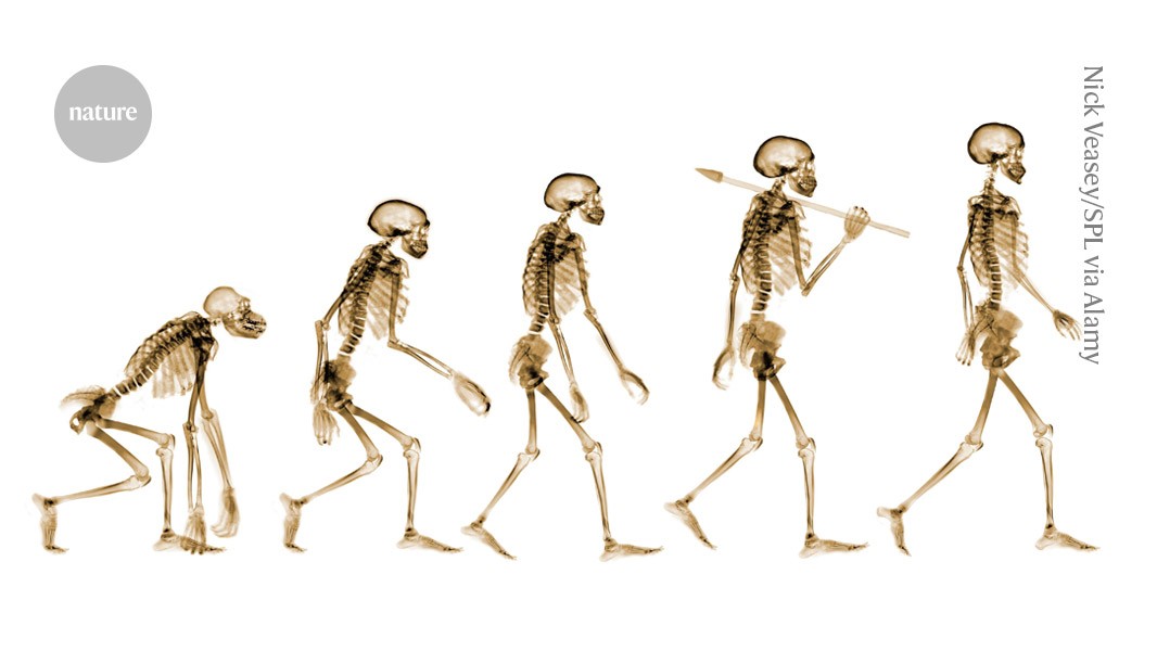
Researchers have identified key structural changes in the development of the human pelvis that enabled upright walking, focusing on the formation and rotation of the ilium during embryonic development, which differentiates humans from other primates and supports bipedalism.
A new tracking technology called LoxCode allows scientists to label and follow individual cells in mouse embryos with unprecedented detail, revealing early lineage biases and asymmetries that influence body part development, with potential applications in medicine and developmental biology.
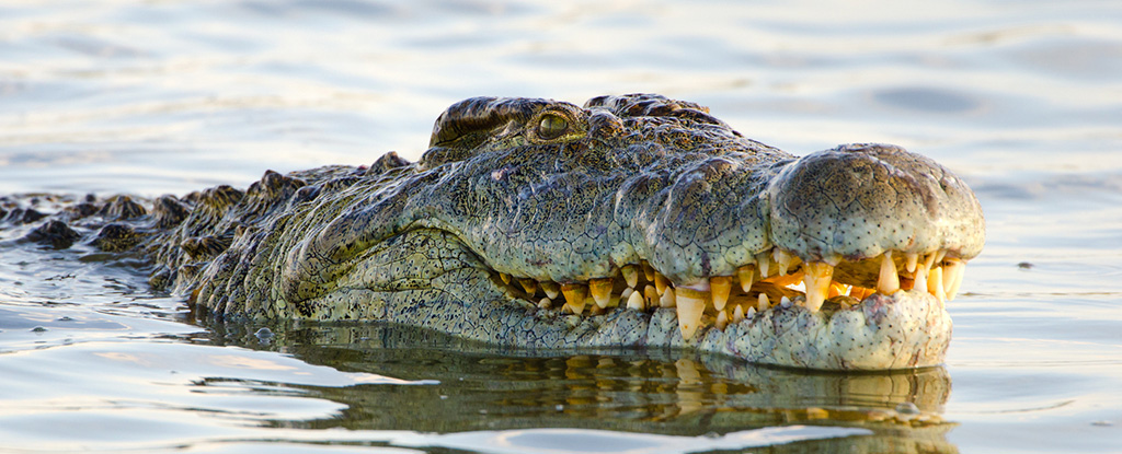
A study by researchers at the University of Geneva has uncovered that the irregular head scales of crocodiles are formed through compressive mechanical instabilities rather than tensile stress. This process occurs during embryonic development, where the skin grows faster than the bone beneath it, leading to the formation of polygonal scales. The findings, published in Nature, suggest that variations in head-scale patterns among crocodilians may be due to evolutionary differences in embryonic skin growth.
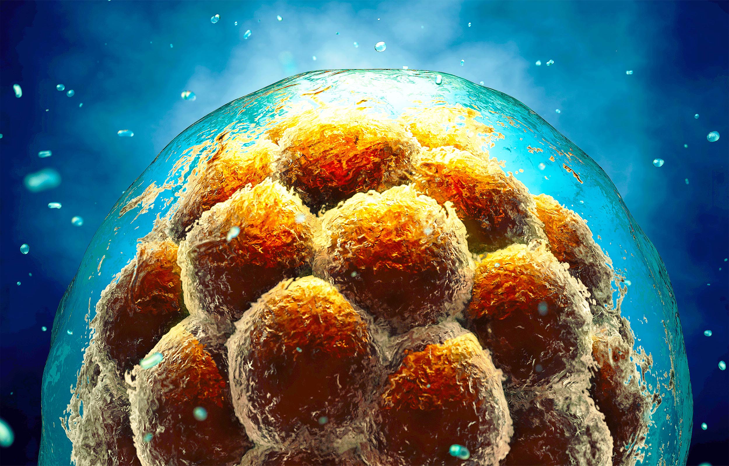
Scientists have discovered that the unicellular Ichthyosporean microbe Chromosphaera perkinsii exhibits embryonic-like cell division, suggesting that the genetic programming for eggs may have existed before animals. This microbe, which has been around for over a billion years, forms a blastula-like cluster of cells, similar to animal embryos. The findings imply that the genetic toolkit for embryonic development was present before the emergence of animals, offering insights into the evolutionary origins of multicellularity. The study, published in Nature, explores whether these similarities are due to a common ancestor or convergent evolution.
Researchers have discovered that the ancient single-celled organism Chromosphaera perkinsii can form multicellular structures similar to early animal embryos, suggesting that the genetic mechanisms for embryonic development may have existed over a billion years ago, before the first animals appeared. This finding, published in Nature, could provide insights into the transition from unicellular to multicellular life and challenge existing views on the evolution of multicellularity.
Researchers from the University of Geneva have discovered that the unicellular organism Chromosphaera perkinsii, which predates animals by over a billion years, forms multicellular structures similar to animal embryos. This suggests that the genetic programs for embryonic development may have existed before animals evolved, or that C. perkinsii independently developed similar processes. The findings, published in Nature, could reshape our understanding of the evolution of multicellularity and embryonic development.
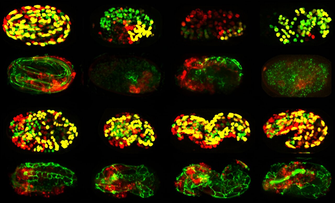
Researchers at UC San Diego have developed a "genetic atlas" using the model organism C. elegans to profile the function of nearly 500 genes during embryonic development. By blocking each gene one at a time and using time-lapse 4D imaging and computer vision, they tracked how these genes influence tissue formation and cell identity. This study, published in Cell, provides new insights into gene functions and their roles in development, with implications for understanding human developmental disorders. The data is now available through an online resource called PhenoBank.
Humans don't have gills because they evolved from fish with lungs, which allowed them to survive on land. Gills need to stay wet to work, making them inefficient for land animals. Early lungs in fish allowed them to gulp air above the surface to supplement oxygen intake. Human embryos have pharyngeal arches resembling gills, which develop into parts of the jaw, throat, and ears. These arches are present in all creatures with heads and are a remnant of early gills.
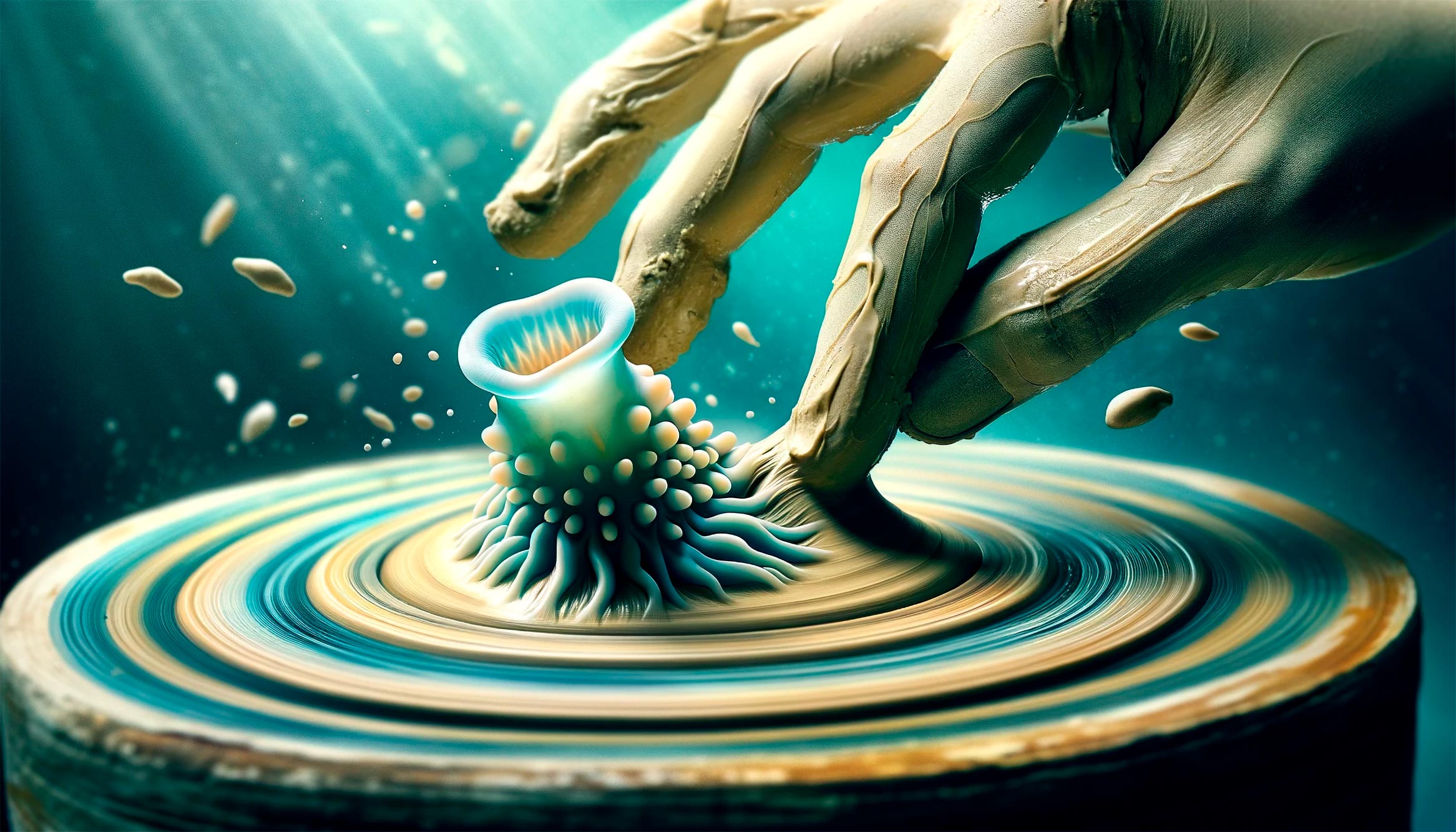
A study from the Institute of Science and Technology Austria reveals that sea squirt oocytes utilize internal friction to undergo developmental changes post-conception, shedding light on the role of friction forces in shaping and forming an evolving organism. Ascidians, or sea squirts, are used as model organisms for understanding vertebrate development due to their similarities with humans. The research provides new insights into the mechanical forces that determine cell and organismal shape, highlighting the pivotal role of friction in embryonic development.
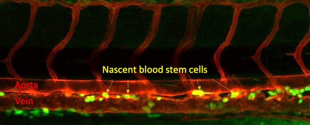
A new study has found that a protein receptor called Nod1, known for its role in recognizing bacterial infections, also plays a crucial role in the development of blood stem cells in embryos. This discovery could lead to the ability to produce blood stem cells from a person's own blood, potentially eliminating the need for bone marrow transplants and improving treatment for leukemia, lymphoma, and anemia patients. The research offers hope for regenerative medicine and could pave the way for creating therapeutic-grade blood stem cells to cure blood disorders.
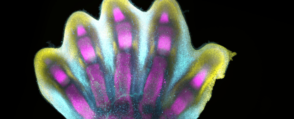
Scientists have created the first human cell atlas of early limb development, revealing in exquisite detail how fingers and toes grow. By analyzing thousands of single cells from donated embryonic tissues, the researchers mapped gene expression patterns and identified distinct cell clusters involved in limb development. They found that the process is highly complex and precisely regulated, resembling a sculptor chiseling away at a block of marble. The study deepens our understanding of how anatomically complex structures form and has implications for research and healthcare. The researchers also showed that limb formation in humans and mice follows similar trajectories, with some differences in activated genes and cell types.