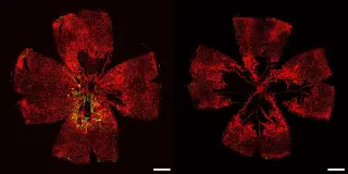
A single-dose gene therapy has successfully repaired damaged retinal connections in adult dogs, offering hope for treating inherited vision loss in humans. The treatment, tested on whippets with a faulty CaBP4 gene, restored vision and neural structures previously thought impossible to repair in mammals.
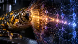
Researchers developed an information-theoretic framework to break a system's local arrow of time into contributions from individuals, pairs, triplets and higher-order groups. When applied to 53 salamander retinal neurons, pairwise interactions accounted for about 66–74% of local irreversibility, and the retina showed a stronger arrow of time under a time-reversible Brownian stimulus than under a natural movie, indicating internal neural processing can generate temporal asymmetry independent of the external input. The approach could be applied to other complex nonequilibrium systems to understand how irreversibility emerges from simple components.
Scientists performed the first human ocular mitochondrial transplantation by injecting mitochondria isolated from a leg biopsy into both eyes of a 26-year-old woman with severe vision loss. The procedure appeared safe with no immune reaction, and post-treatment results included temporary improvements in pupil light responses and evidence of organized visual-cortex activity, though there was no restoration of visual acuity. The study is a single-patient, uncontrolled case, so causation cannot be established, but it suggests a potential for repeated mitochondrial dosing and warrants further, controlled trials to explore whether healthy mitochondria can energize damaged retinal cells.
Experts warn that looking at the sun during the eclipse can cause solar retinopathy, with central vision distortion, a grey/yellow/dark spot, or increased light sensitivity that may appear hours later; other signs include eye pain, redness, grittiness, or sudden vision changes. Because the retina has no pain receptors, damage can go unnoticed initially. Only specially designed eclipse glasses protect against this; if you notice any vision change, seek urgent assessment from an optician or eye unit for retinal scans and appropriate care. The UK saw fewer cases than in 1999, but precautions remain essential.
A Staten Island woman looked at the 2017 eclipse without proper eye protection for about 20 seconds and soon developed a crescent-shaped blind spot in her vision. High-resolution retinal imaging then showed a matching crescent-shaped patch of photoreceptor loss, directly linking the sun’s crescent to the damage; solar retinopathy is caused by irreversible photoreceptor death with no available treatment, underscoring the dangers of viewing an eclipse unprotected.
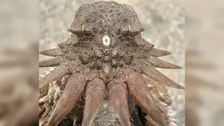
A new evolutionary model proposes that vertebrate eyes, including humans', arose not from scratch but by repurposing components from a single ancient median eye about 600 million years ago. Over time, this median organ diversified into left/right retinas and the pineal system, with embryonic retina development tracing back to brain tissue. The scenario helps explain how different cell lineages fused into the vertebrate visual circuitry, supported by comparative data across species and recent studies of early retinal cell types.
Duke University researchers used induced pluripotent stem cells (iPSCs) to grow retinal endothelial cells—the vessels lining the retina—and demonstrated they can form functional vascular networks in lab conditions. When injected into mouse models of retinal disease, the lab-grown cells integrated with existing tissue to rebuild blood vessels and restore retinal function, suggesting a cheaper, scalable path toward preventative therapies for conditions like diabetic retinopathy.
Eye health can reflect brain health, and untreated vision problems are a major, modifiable dementia risk factor. While vision loss doesn’t directly cause dementia, correcting vision (updating lenses, cataract/AMD treatment) can help keep the brain engaged with the visual world and may reduce dementia risk by up to about 30%. Eye exams and retinal imaging (OCT) might even help track dementia progression. Regular eye care, addressing underlying eye diseases, avoiding smoking, and maintaining vision health can support cognitive function, even after a dementia diagnosis.
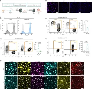
Researchers derived retinal endothelial cells from human iPSCs via the Norrin–Fz4–Wnt/β-catenin axis, enhanced barrier maturation with RepSox, and demonstrated retinal-specific markers, GLUT1/p‑gp transporter activity, and robust, lumenized 3D networks. In vivo, iRECs integrated into ischemic mouse retinas, promoting revascularization, restoring normal vascular morphology, and reducing permeability in an oxygen-induced retinopathy model. In vitro, iRECs formed functional iBRB microphysiological systems with retinal pericytes (iRPCs), recapitulating iBRB structure and barrier function. Transcriptomics confirmed retinal gene signatures aligning with primary RECs, though not fully matching HRECs. The work establishes iRECs as a scalable, tissue-specific cell source for retinal disease modeling (notably diabetic retinopathy) and therapeutic cell-based strategies, while acknowledging limitations in biomarker breadth and complex pericyte identity for future refinement.
Astronauts aboard the International Space Station report seeing flashes of light with eyes closed caused by high-energy cosmic rays passing through their bodies, either directly ionizing the retina or via Cherenkov radiation. Historical experiments (SilEye/ALTEA) linked particle tracks to the flashes, confirming the phenomenon and highlighting space radiation as a detectable, personal readout of exposure and a major shielding challenge for long-duration missions.
Researchers analyzed 62,876 retinal photos from more than 40,000 UK Biobank participants with deep learning to predict 12 Alzheimer's-related risk factors. They found retinal features like vascular stiffening, reduced vessel density, and thinning of the optic nerve linked to later risk, suggesting retinal imaging could flag at-risk individuals years before symptoms (though it cannot diagnose the disease). The study highlights retina as an integrated biomarker of cumulative risk and was published in the Journal of Alzheimer's Disease.
Life Biosciences has dosed the first patient in an FDA-cleared trial of ER-100, a gene-therapy approach to reverse age-related degeneration of retinal neurons in glaucoma by reprogramming cells with Yamanaka factors activated by doxycycline for eight weeks. The work, building on Harvard's David Sinclair research, aims to rejuvenate vision but faces expert caution about potential runaway cell growth or cancer from cellular reprogramming.
Researchers in South Korea developed experimental smart contact lenses with tiny electrodes that deliver retinal electrical signals using temporal interference to target mood-related brain circuits in mice, improving depression-like behavior in animals with damaged retinas; the approach is early-stage, not tested in healthy retinas, and many challenges remain before any human use.
A 2026 Nature Communications study shows Greenland sharks can live for centuries and retain a functional visual system, overturning the long-standing claim that they are nearly blind due to an eye parasite. Earlier radiocarbon dating suggested lifespans up to ~392 years, but the new work finds preserved retinal tissue and active low-light vision, prompting a rethink of how we understand ultra-long-lived deep-sea animals.
Population studies in Singapore and the UK found that markers of faster retinal aging are associated with lower bone density and higher osteoporosis risk, suggesting retinal images could serve as a noninvasive screening tool, with AI models like RetiAGE predicting future fractures.